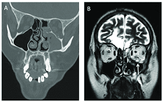
In the wake of the ongoing Coronavirus Disease 2019 (COVID-19) pandemic, a new epidemic of COVID associated mucormycosis (CAM) emerged in India.
Early diagnosis and prompt treatment of this deadly disease are of paramount importance in improving patient survival. MRI is the cornerstone of diagnosis of early extrasinus disease, particularly intracranial complications which have traditionally been associated with a high mortality rate.
Sehgal et al from Maulana Azad Medical College and Lok Nayak Hospital, New Delhi India ; presented the sinonasal, perisinus, orbital and intracranial involvement in CAM. The aim of the review is to familiarise the reader with the MR imaging spectrum of CAM with special focus on intracranial complications and a brief account of their impact on patient prognosis in the author's experience.
Special emphasis is made on intracranial disease which is categorized into vascular, parenchymal, meningeal, bony involvement and perineural spread. Vascular complications are the most common form of intracranial involvement. Some unusual yet interesting imaging findings such as nerve abscesses involving the optic, trigeminal and mandibular nerves and long segment vasculitis of the internal carotid artery extending till its cervical segment are also illustrated. In our experience, patient outcome in CAM (survival rate of 88.5%) was better compared to the pre-pandemic era. Presence of intracranial disease also did not affect prognosis as poorly as traditionally expected (survival rate of 82.8%). Involvement of brain parenchyma was the only subset of intracranial involvement that was associated with higher mortality (p value 0.016).


No comments:
Post a Comment